- World's Leading Trade FairLASER World of PHOTONICSJun. 24-27, 2025
- Parallel eventsWorld of Photonics CongressJun. 22-27, 2025
- Trade fair networkChinaMar. 11-13, 2025
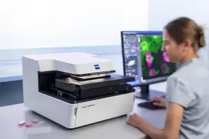
Being able to monitor processes in living cells for hours or even days is a dream researchers have had for a long time. But the very intensive laser light needed to excite fluorescence damaged the cells and influenced the processes. A team from ZEISS has now solved the problem and received the “Deutscher Zukunfts–preis” at the end of October in recognition of their achievement.
The potential of the new microscopy method is already becoming apparent. For example, a team of malaria researchers was able to observe the life cycle of the pathogens in detail using the award-winning ZEISS Lattice Lightsheet 7 microscopy system. They also identified proteins that can be targeted by new drug and therapy concepts. Research into infectious diseases and cancer is also being put on a completely new footing thanks to an insight with high temporal and spatial resolution into processes in living cells lasting hours or even days. After all, it is now possible to see in detail how cells react to specific active substances or what happens when they are hijacked by viruses or bacteria. According to Dr. Thomas Kalkbrenner, the spokesman for the ZEISS team that won the award, the new process also has potential in situations where so-called organoids are used. The miniature organs cultivated in Petri dishes are used in drug research to test the effects and side effects of active substances. In the future, organoid testing could make the majority of animal testing obsolete. However, this will only be possible if the light-sensitive organoids are not damaged by the laser light hitting them and their behavior is not influenced. The problem is that the laser radiation used is 1,000 times more intense than the sun’s radiation.
So how exactly did the research team led by Kalkbrenner and his colleagues Dr. Jörg Siebenmorgen and Ralf Wolleschensky go about shielding the living cells from the influence of the intense laser light that is essential for fluorescence excitation? Instead of exposing the cell structures directly, they feed the light as a light sheet laterally perpendicular to the detection lens. This strategy reduces the intensity of the radiation penetrating the tissue. To generate the very thin yet long light sheets needed for cell observation, the team resorted to concepts of lattice-shaped lights sheets generated by a spatial light modulator (SLM). Scanners dither the lattice structure to create a smooth light sheet which is then projected onto the sample through exactly the observed portion.
With this, however, the team had only overcome the first hurdle. Because cells are grown in laboratories around the globe in standardized Petri dishes or on multiwell plates, the ZEISS team had to make 3D long-term cell observation possible through their glass bottoms. To make this possible with the lattice light sheet strategy and the side-fed laser, the team had to tilt the entire arrangement, which greatly distorted the microscopic view through the glass bottoms. In more technical terms, variations in refractive index occur: because fluorescence is emitted from the sample, it passes through aqueous cell culture media, the tilted glass coverslip and a water immersion before reaching the detection lens. The developers also solved this optical problem. By integrating free-form optics into the detection beam path, they were able to compensate for the distortions and even the manufacturing-related shape deviations in the Petri dishes and multiwell plates.
The team from ZEISS Microscopy was able to integrate the highly complex process into an easy-to-use, compact system with a high level of automation. In recognition of this, it received the Deutscher Zukunftspreis 2022 at the end of October. Following the award for the development of EUV lithography in 2020, this is already the second ZEISS team in this decade to receive the €250,000 prize from German President Frank-Walter Steinmeier. Speaking at the award ceremony, Executive Board member Dr. Jochen Peter said the ZEISS Group was proud of the team and its extraordinary achievement in developing the ZEISS Lattice Lightsheet 7. “At the same time, the award is confirmation of our company’s innovative strength which encourages both economic and social progress,” he said.
In the video, which is unfortunately only available in German, Dr. Thomas Kalkbrenner, Team Leader and Lead Architect R&D Special 3-D at Carl Zeiss Microscopy, explains the challenges the team overcame and the technological and social potential the ZEISS Lattice Lightsheet 7opens up for biological and medical research.