To exploit AI potential, we need photonomics
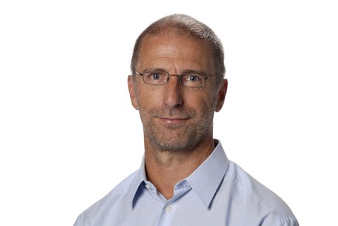
As Chairs of the European Conferences on Biomedical Optics (ECBO) and their sub-conference “Translational Biophotonics: Diagnostics and Therapeutics,” Prof. Ronald Sroka and Prof. Lothar Lilge have provided current insights into technological trends and challenges in biophotonics. These are underpinned by Prof. Sroka’s professional experience as Scientific Head of the Laser Research Lab at the University Hospital of Munich, and by Prof. Lilge’s research at the University of Toronto’s Princess Margaret Cancer Centre as part of Canada’s University Health Network. In a joint interview, they both speak about new diagnostic procedures ready to make the transition to clinical practice, about what they would like from laser manufacturers and politicians, and about the void standing in the way of the productive use of machine learning and artificial intelligence (AI).
Looking back at ECBO 2023, what are your thoughts?
Prof. Ronald Sroka: After the four-year forced break due to the pandemic, it was great to be able to meet up again in Munich and talk to each other face to face. ECBO alone had six sub-conferences and was just one of the five trade conferences at the World of Photonics Congress, at which more than 3,600 scientists from all over the world presented the latest research approaches and results this time in specialist presentations.
Prof. Lothar Lilge: The biggest sub-conference at ECBO was “Translational Biophotonics: Diagnostics and Therapeutics,” chaired by myself and Zhiwei Huang of the National University of Singapore. When we first launched this conference, it was the smallest ECBO conference with just 40 submissions. This year we had almost 120 presentations, which clearly shows that biophotonics is on its way out of the research lab and into clinical practice. Funding bodies from all over the world are now expecting their investments over the past 20 years to get ready for the market.

Your conference is about biophotonic diagnostics and therapy. In which area do you see greater momentum?
Lilge: Most definitely in diagnostic applications. They account for 85 to 90 percent. In cases where photonic technologies are used for therapeutic purposes, we are seeing a trend toward weaker light sources.
Sroka: The long-discussed translational effect has now set in: More and more photonic processes are finding their way into medical practice. But what does concern us with regard to that is the Medical Device Regulation in the EU. Now, of all times, the regulation is stifling the optimistic mood and causing congestion at the interface between research and application. Many companies are shying away from the effort involved in the approval of medical devices. At the same there is also great restraint on the clinical side to trial devices that have not yet been certified in pilot applications and studies. But without their clinical trials, the approval procedure for the new approaches won’t get going at all. This knot needs to be untangled.
Which biophotonic methods are on the on the brink of being launched on the market, and what are their benefits?
Lilge: As light has proven to be a highly versatile and valuable tool in diagnostics, there are quite a lot at the moment, starting with endoscopy, which allows almost every area of the human body to be examined thanks to increasing miniaturization. The outside diameter of some modern endoscopes is now only 1.2 millimeters ...
Sroka: … and they are increasingly coupled with multimodal imaging processes, enabling whole new insights into vessels and deep tissue layers. In line with that there is a clear trend toward imaging without adding markers, whether it’s via optical coherence tomography (OCT), with Raman spectroscopy, multispectral and hyperspectral imaging, or also with the aid of photoacoustics that innervate the targeted tissue with short light pulses and measure its reaction by ultrasound. The method combines the selectivity of the light with the penetration depth of the ultrasound, hence delivering high-resolution three-dimensional insights better than conventional ultrasonic imaging. Combining OCT with endoscopy extends the OCT application spectrum from skin and eye examinations into the depths of the body. That adds a new high-precision method to the toolbox of minimally invasive diagnostics.
Lilge: Talking of hyperspectral imaging, increasingly broader spectral ranges are being used for diagnostics. At the same time, however, there is also a trend toward methods that are based on analyses of the wave character of light and, for example, make use of polarization for that. We are getting better and better at understanding the interaction of circular and linear polarized light – and can, for example, close in very accurately on the irradiated tissue and the penetration depth of the light via clockwise or counterclockwise reflection.
How do these applications change the requirements of laser beam sources?
Lilge: Current laser beam sources are reaching their limits with hyperspectral imaging. In white light applications, in particular, the brightness of the light often leaves much to be desired – and the individual wavelengths influence one another in their interaction with the tissue. New practical solutions that emit different, individually isolated wavelengths would be required for clinical use. It should be possible to control them individually and combine them, as required. Systems that enable wavelengths to be switched very quickly, but not continuously are also conceivable. That would help with hyperspectral imaging to achieve clearer diagnoses through increased brightness in defined wavelength ranges. New light sources are also needed for polarization-based imaging. It would be very helpful if there were light sources with directly programmable polarization direction and also directly electronically controllable solutions for detection. There is a need for development both with regard to the light sources and in the area of polarization filtering and the receptor and analysis tools. I also see the potential here of a combination with endoscopes and other imaging devices here.
Such solutions would be very helpful for the early detection of conspicuously deviating tissue structures that are often the precursors of tumors. Being able to perform minimally invasive diagnosis of such structures without removing tissue would be fantastic progress.
Sroka: We are getting closer and closer to the "optical biopsy" with the developments to date and those outlined above. Various research teams are also working on the potential of polarization and depolarization processes for analyses at cellular and vascular level. The focus is on parameters such as elasticity and permeability, whose monitoring may also hold a key to identifying resistances.
What is the situation with beam sources for photoacoustics?
Lilge: As already mentioned, this is about insights into deep tissue. With polarization and OCT applications, it has so far mostly been about the analysis of tissue surfaces. More powerful light sources with different, individually controllable wavelengths are required to examine deeper layers with the aid of photoacoustics or also with diffuse optical spectroscopy. Only then can larger tissue areas be examined in everyday clinical practice. Different wavelengths are needed, for example, to be able to differentiate between arterial and venous blood using photoacoustics. This analysis becomes interesting when it’s combined with structure analyses of surrounding tissue on the basis of its fluorescence. This can be done with contrast agents. But that requires the already mentioned further development of the light sources – toward more brightness and more individually controllable pulses. These should not, however, be as complex as an optical parametric oscillator (OPO), as there are usually no physicists available in the clinics to operate the devices. Practical, easy-to-use solutions are needed. Units with selectively controllable, variable level laser diodes, which achieve a high performance in their respective wavelength, would be conceivable here. Instead of having to couple numerous different lasers, systems that deliver different wavelengths from one source would be ideal. Because we could then apply them immediately in endoscopy and other intravital procedures
Sroka: We had our own session on the possibilities of “Diffuse Optical Spectroscopy and Imaging”. This is also a very exciting field of innovation. Among other things, it’s about the optical detection of tissue perfusion. There are a variety of processes for that, some integrated into wearables for continuous health monitoring. That can be very useful, be it in sport, home care or also for monitoring the efficacy of drug therapies. I also see big potential for early diagnostics, since anomalies are detected sooner with continuous recording of vital data. And then there are also spectacular research approaches, such as optical, non-invasive monitoring of neuronal processes in the brain – with light through the skullcap.
Many presentations at ECBO 2023 dealt with the use of deep learning and AI. Where does biophotonics stand on this issue?
Lilge: Despite all the optimism surrounding the potential of these technologies, I still see a large gap here that needs to be filled. In biology, genomics, proteomics or metabolomics have laid the basis, both at data and cognitive level, for AI and deep learning to be used highly productively today. That’s precisely what is lacking in photonics. We would need photonomics to both set up a shared public access database on a global scale and to develop common methodical understanding. AI offers huge potential for biophotonic diagnostics and therapy. That also applies to interpreting light signals and to adapting photonics-based therapy procedures to individual patients.
Sroka: The use of AI and deep learning was practically omnipresent in the ECBO presentations. With appropriately trained algorithms it promises much potential to unburden doctors, since they then only have to look at a fraction of the image data in which the AI finds anomalies. However, the database required for that is really too thin, so the AI support lacks substantive content in many cases. Until algorithms in tissue images can identify a previously undetected tumor, they must be trained using very big data volumes. But we don’t have those. Added to that is the problem of insufficient comparability of images from the different manufacturers’ devices. Standards are missing. Researchers are currently endeavoring to solve this problem with software: The goal is to objectify image data and make it comparable using software. Reference systems like these are essential for this, so that we can train the algorithms with as broad a database as possible. Red must be red, with exactly the same color values. There is a great need for development, and the interoperability of the procedures and devices of different manufacturers must be established.
Lilge: A consortium, ideally international, is required, which would commit to setting up a publicly accessible database and ensure its setup at political, legal, clinical and practical level. After all, it’s also possible in genomics. Anonymizing datasets and ruling out misuse is not rocket science. It can be resolved with binding sets of rules and regulations, which should have been in place long ago! To date, we’ve been relying on local cooperation. Our institute shares anonymized cancer diagnosis image data, for example, with partners. But this won’t require hundreds or thousands of data records – given the diversity of clinical pictures and the variance of their manifestations, hundreds of thousands of them will be required to train the algorithms in the necessary breadth and depth. And Ronald is right: We also need clear standards in imaging. When every manufacturer retains their own proprietary solutions, then the comparability of the data suffers. Compare it with the evolution of a language. The more dialects that are integrated, the more complicated and incomprehensible it becomes for all involved.

